Abrasion: An area damaged by scraping.
Injury to superficial skin epithelium due to sliding force, compression or pressure; i.e. scrape
- Patterned abrasion: injury in which a pattern is transferred from the impacting object or intermediary material (i.e. clothing); can be used to identify weapon
- Road rash is an abrasion caused by the road surface; commonly seen in pedestrian-motor vehicle accidents (MVA) or bicycle accidents
- Antemortem abrasions are usually reddish brown; postmortem abrasions are yellow or transparent (due to absence of blood flow). Link.
Axillary: Relating to the armpit or corresponding part.
Dorsum: The dorsal (back) of a structure.
Intercostal: Situated between the ribs.
Interphalangeal: Occurring between the phalanges (bones of fingers and toes).
Laceration: A tear in the skin or flesh.
Although emergency medicine providers commonly describe any break in the skin as a laceration, this terminology is forensically and technically incorrect. A laceration is defined as a tear in tissue caused by a shearing or crushing force. Therefore, a laceration is the result of a blunt-trauma mechanism. A laceration is further characterized by incomplete separation of stronger tissue elements, such as blood vessels and nerves. These stronger tissue elements account for “tissue bridging” which is seen in lacerations. In addition, lacerations commonly occur over bony prominences and tend to be irregularly shaped with abraded or contused margins. Lacerations are typically caused by hard objects like a pipe, rock, or the ground. An easy way to remember the difference is to think of a glass beer bottle. If someone takes the bottle and smashes it over someone’s head and the skin is opened, that is a laceration. If a person breaks the bottle on a table and uses the piece to slash someone, it is an incised wound. Link.
Medially: Situated or pertaining to the middle.
Oblique: Neither parallel or at a right angle.
Petechia: A small red or purple spot caused by bleeding into the skin.
Subcutaneous: Situated or applied under the skin.
Thoracic: Relating to the thorax (cavity enclosed by the ribs), or between neck and abdomen.
Transected: Cut across or make transverse section in.
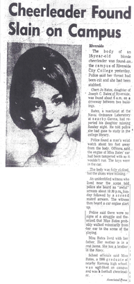
31st October 1966
07:15: Called at home by Chief Deputy Coroner William J. Dykes about the possibility of a homicide.
08:30: Called at office by Chief Deputy Coroner William J. Dykes to proceed to 3680 Terracina Street, Riverside, California.
09:05: Arrived at scene. Body in capris, white sandals without socks or hose and loose pick moderately heavy blouse. Laying mainly face down.
09:24: Body examined at site; skin surface cool, rigor present in all extremities but more so in lower than superior members.
09:31: Liver temperatures 26 and 28 degrees celsius.
10:50: I helped remove clothes.
11:00: Autopsy started and several hairs removed from base of right thumb and placed in a 4 by 1 1/2 inch clear plastic container held by Detective Earl T. Brown. I removed blood from the heart. Subsequently placed in the laboratory refrigerator.
13:42: Autopsy completed.
1 November 1966: Vaginal swab washing sediment smears made; no spermatazoa identified.
2 November 1966: Blood typed AB; Rh. (D) positive.
3 November 1966: Tests for barbiturates, narcotics and ethanol - negative.
[1] Green eyes and brown scalp hair.
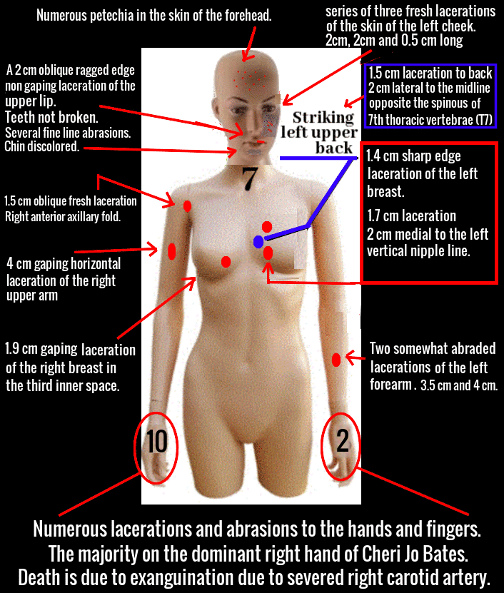
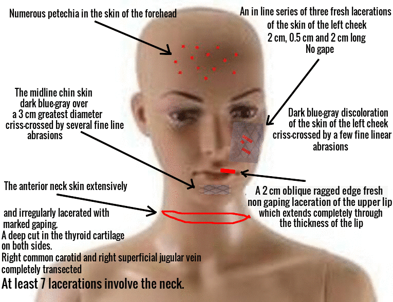
[2] A 2 cm oblique ragged edge fresh non gaping laceration of the upper lip on the left side, that angles laterally from above and extends completely through the thickness of the lip. The teeth behind are not loose or broken.
[3] A dark blue-gray slightly swollen discoloration of mainly the mucocutanous portions of the upper and lower lips of the right side, involving a 2 cm greatest diameter.
[4] Numerous petechia in the skin of the forehead.
[5] An in line series of three fresh lacerations of the skin of the left cheek, angling from above in front slightly downward and posteriorly. The anterior is 2 cm long, the intermediate 0.5 cm long and the posterior one 2 cm long. The overall length is about 3 cm and all extend into the superficial subcutaneous tissue, but do not gape.
[6] An area of dark blue-gray discoloration of the skin of the left cheek and chin involving mainly the anterior two of the lacerations just mentioned. It is also criss-crossed by a few fine linear abrasions.
[7] The midline chin skin dark blue-gray over a 3 cm greatest diameter and criss-crossed by several fine line abrasions.
[8] The anterior neck skin extensively and irregularly lacerated with marked gaping. A deep cut in the thyroid cartilage on both sides and the right common carotid is completely transected as well as the right superficial jugular vein.
[9] A gaping, about 1.5 cm oblique fresh laceration of the skin of the right anterior axillary fold, centered 5 cm from the apex produced by the anterior axillary fold and the right arm. It is probed into the subcutaneous tissue about 1 cm.
[10] A 1.4 cm fresh vertical gaping sharp edge laceration in the upper medial quadrant of the left breast in the second ICB (Intercostal block).
[11] A 1.9 cm gaping sharp edge fresh laceration of the lower medial quadrant of the right breast in the third inner space.
[12] A 1.7 cm mainly transverse fresh laceration of the skin of the left chest over the 5th rib and centered about 2 cm medial to the left vertical nipple line.
[13] A 4 cm gaping fresh sharp edge, mainly horizontal laceration of the right upper arm anterior and medially that extends through the fat and into the muscle.
[14] A 1.5 cm greatest diameter fresh abrasion with dark blue discoloration of the skin at the base of the dorsum of the right thumb.
[15] A few light linear criss-crossing abrasions of the skin of the dorsum of the right hand involving mainly the medial half over the metacarpals.
[16] Two abrasions, each about 1 cm in greatest diameter, in the skin at the base of the dorsum of the right middle finger.
[17] A 2-3 mm fresh abrasion of the skin of the dorsum of the middle of the right 4th finger.
[18] Considerable partially dried blood over the hand and especially about the fingers and under the unpainted moderately long (2-3 mm) but not carefully manicured fingernails.
[19] Two fresh 3-5 mm greatest diameter abrasions in the skin over the 2nd IP (interphalangeal) joint of the lateral aspect of the right index finger.
[20] An irregular 1.3 cm greatest diameter recent abrasion of the skin over the 2nd IP joint of the medial aspect of the right little finger.
[21] A more or less AP interrupted fine abrasion type laceration in the skin of the base of the medial aspect of the right little finger.
[22] A curved and interrupted moderately deep laceration (2 cm overall} in the skin of the base of the lateral aspect of the right index finger.
[23] Two somewhat abraded fresh lacerations of the skin of the volar surface of the left forearm more or less in the mid portion. They run from lateral to medial, the longer is 4 cm and the shorter is 3.5 cm and more lateral than the former. These extend into the subcutaneous tissue.
[24] A more or less Y shape laceration in the skin of the dorsum of the left hand medially and at the junction of the wrist and hand.
[25] An irregular laceration of the skin of the dorsum of the left hand in line with the middle finger and is about the mid area.
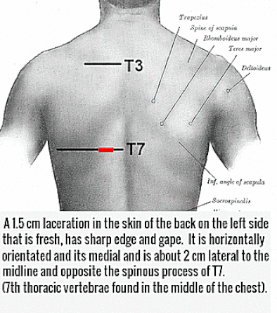
[26] A 1.5 cm laceration in the skin of the back on the left side that is fresh, has sharp edge and gape. It is horizontally orientated and its medial and is about 2 cm lateral to the midline and opposite the spinous process of T7 (7th thoracic vertebrae found in the middle of the chest).
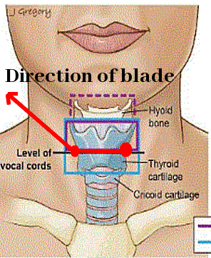
The thyroid cartilage is obliquely cut on each side. Each cut is about 3 cm long and angles from lateral above to medially below forming a somewhat Y shape structure with the apex at about the mid portion of the thyroid cartilage anteriorly. The upper edges are cut much deeper than the inferior portions. The medial edges of the upper portions of the right and left lobes of the thyroid are partially incised. The right and left superficial jugular veins are completely transected. The right common carotid artery is completely transected about 2 cm above its origin.
Cardiovascular System:
The about 200 gram heart is in a pericardial sac that has no lesion of either surface or the space. There is no significant coronary artery disease, congenital anomaly, endocardial fibroelastosis or significant dilatios.
Respiratory System:
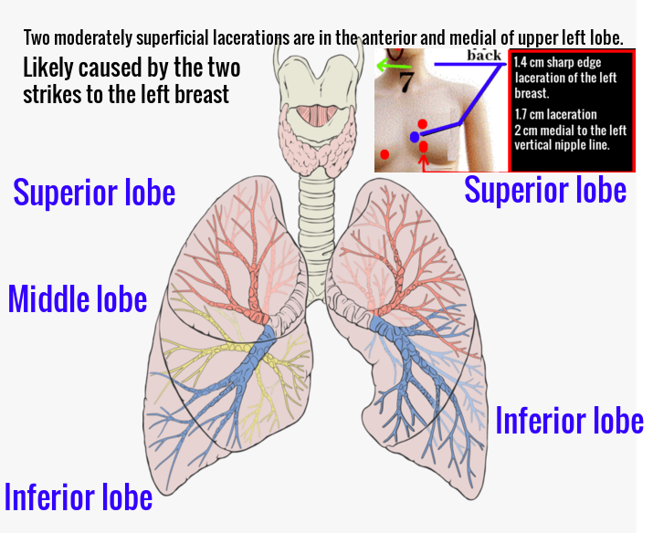
The about 400 gram lung is free in its pleural sac and has no lesion of its surface or interior. The about 400 gram left lung is also free in its pleural space. Two moderately superficial lacerations are in the anterior and medial of its upper lobe. There is no significant foreign body obstruction of the trachobronchial tree, pulmonary embola significant atelectasis (collapse of lung tissue with loss of volume), emphysema, pneumonia, significant hemorrhage into the lining of the larynx or about the other soft tissues about the larynx.
Gastrointestinal System:
There are no lesions of the peritoneum, omastum or mesentery. There are no lesions of the oesophagus. The stomach contains at least 100 millilitres of thick food with particulate food particles in which are easily recognized reasonably large pieces of apparently beef along with vegetable particles being either and/or celery and onion and white curd like particles floating in the gastric contents that appear to be either milk or cottage cheese. There is no lesion of the stomach, duodenum, remaining small bowel or large bowel including the rectum. The 120 gram pancreas is in its usual position and has no lesion of its surface or interior.
Hepatic System:
The about 1250 gram liver is in its usual position and has no lesion of its surface or interior. A gallbladder is present with no lesion of the wall or lumen including calculi and there are no lesions of the remaining extrahepatic biliary duct system.
Lymphocytic System:
The about 90 gram spleen is in its usual position and has no lesion of its surface or interior. There are no significant lymph node changes in the thorax or abdomen.
Urinary System:
Each about 160 gram kidney has a capsule that strips easily leaving a smooth cortical surface with no lesion. The interiors have the usual cortical and medullary markings with no lesions and there are no lesions of the calyceal systems, renal pelvis, urethra or urinal bladder.
Reproductive System:
The external genitalia are those of an adult female. The vaginal orifice has no hymen but the usual ring of carcaculae are present. There is no significant amount of fluid in the vaginal canal, however, dry cotton swabs are taken. The uterus, ovaries and oviducts are present and there is no evidence of pregnancy.
Lacerations of the about 20 gram thyroid have been previously mentioned. There are no other lesions of its surface or interior. Each adrenal is in its usual position, has no lesion of its surface or interior and they total about 11 grams.
Skeletal and Muscular Systems:
There is a 1.5 cm oblique cut in the bony portion of the 5th rib anteriorly on the left corresponding somewhat to the skin lacerations over it. There are no other bony lesions and fractures are looked for. The lacerations of skeletal and muscle have been mentioned under specific areas.
[26] A 1.5 cm laceration in the skin of the back on the left side that is fresh, has sharp edge and gape. It is horizontally orientated and its medial and is about 2 cm lateral to the midline and opposite the spinous process of T7 (7th thoracic vertebrae found in the middle of the chest). See picture above.
Lacerations of face, neck, hands, arm, forearms, lung, common carotid artery, thyroid and thyroid cartilage.
Abrasions of face, hands and forearms. Petichiae of forehead.
Death Certificate Diagnosis:
Hemorrhage, due to laceration of right carotid artery.
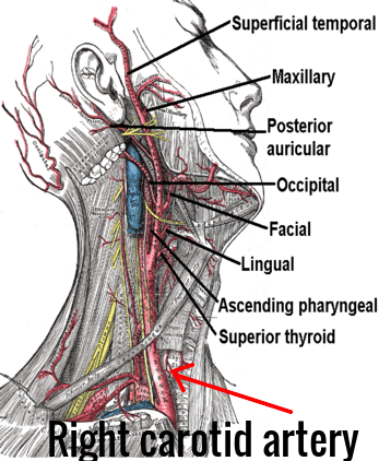
Death is due to exanguination due to severed right carotid artery due to laceration of neck due to probable knife. As the result of the rapid blood loss the subject would go into shock rapidly and undoubtedly would become unconscious and die in several minutes. The time interval of minutes might make one wonder why she wouldn't scream and run for help, however, it is very likely that the suddenness and fear caused by the attack caused her to faint, hence, allowing for many long and loud calls for help. The syncope would then blend into the unconsciousness from blood loss and into death.
The state of rigor mortis, post mortem lividity and body temperature at 09:23, 31st October 1966 indicated she had been dead between 9 and 12 hours.
The lacerations are typical of knife cuts and the minimum instrument dimensions for a knife blade would be half inch wide and three and a half inches long. There were no "tell tale" marks to indicate it had been plunged in "up to the hilt". The position(s) of the assailant is/are not "proof positive" from the study of the body.
The gastric contents suggest she had eaten a supper type meal probably not more than 2-4 hours before her death.
Vaginal contents for spermatazoa, spermine and choline.
At least 7 lacerations involve the neck.
The right common carotid artery is transected only once.
The sequence of the lacerations of the neck cannot be absolutely established nor is there any reasonable suggestion of their order of occurrence from the examination of the body.


 RSS Feed


